(Click on image for larger view.)
(Click on image to view video as MOV file, or as MPEG-1 file.)
(Click for larger view.)
April 8, 2007
Microscope design: Rudolf Oldenbourg and Marc Levoy
Software and web page: Zhengyun Zhang
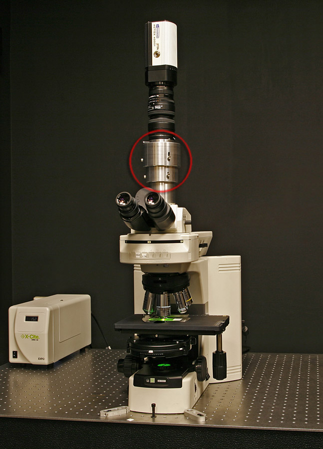
At left below is a prototype light field microscope (LFM) assembly that is portable, inexpensive (about $2,000, not including the base microscope), and can be installed on a compatible microscope in less than a minute. In this example the base microscope is a Nikon Eclipse 80i. We have also built a version for Zeiss microscopes. Attached to the Nikon's camera port (above the eyepieces) is a custom-machined housing for our microlens array (circled in red). The array contains 288 x 192 microlenses, with each microlens measuring 125 microns on a side. We image the microlenses through a relay lens system consisting of two Nikon f/1.4 50mm lenses. The camera (at top) is a Retiga 4000R 2048x2048 cooled CCD camera.
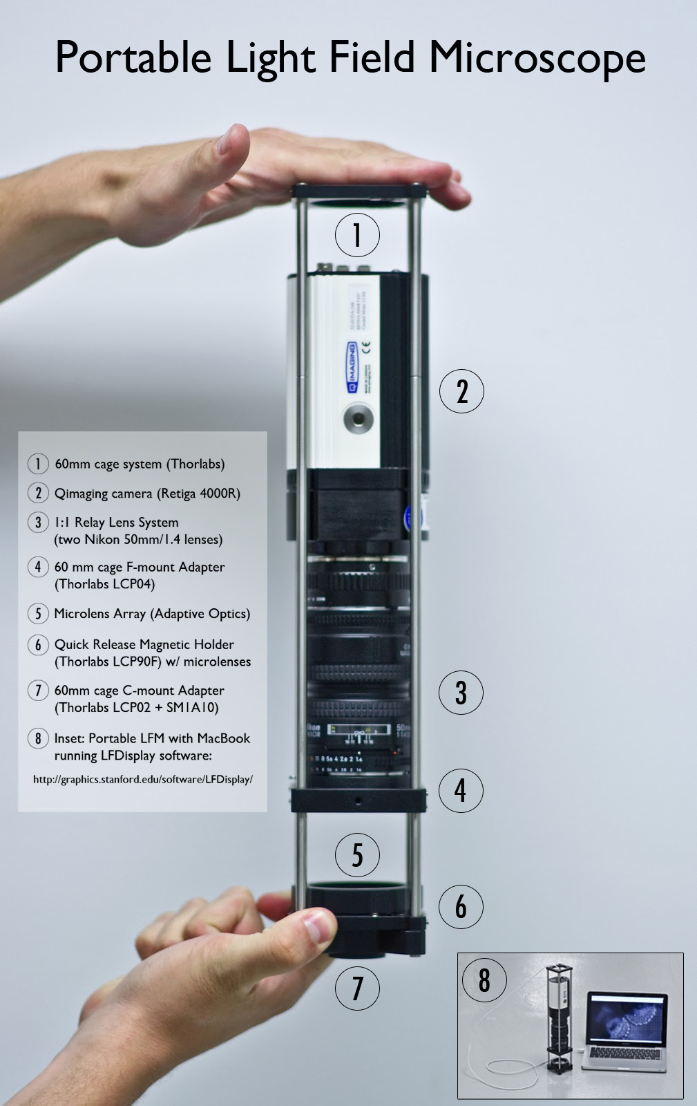
Note added June 2010: At right below is another version of the light field microscope, designed by Logan Grosenick and Todd Anderson in 2010. It mounts to a C-mount camera adapter, which means it can be fitted to the camera port of any research-grade microscope, using a standard adapter that can be purchased from the microscope vendor. The rigid frame makes it easier to remove and re-install the assembly on a microscope without affecting its alignment. This design also simplifies the swapping of microlens arrays. (Different arrays are needed for different microscope objectives, as described in this Practical Introduction to Light Field Microscopy, written by Zhengyun Zhang.)
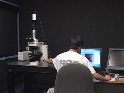
At center below, PhD student Zhengyun Zhang uses both hands to manipulate light field micrographs using his software viewer. With his left hand, he is translating the microscope stage in the usual way. With his right hand, he is digitally tilting the specimen around the X or Y axes using a mouse. Since light fields are being continuously streamed to his software from the Retiga camera, and his software can compute oblique or refocused views from these light fields at roughly 30 Hz, the view he sees on the screen is both interactive and live. This software runs on any PC with a reasonably fast graphics accelerator card. The software is called LFDisplay; click on the name to download it. Several sample light fields are included in the download. Also included is a "protocol" (i.e. step-by-step instructions) on how to build a Light Field Microscope (LFM).
|
Light Field Microscope
(Click on image for larger view.) |
Real-time Viewer
(Click on image to view video as MOV file, or as MPEG-1 file.) |
Alternative design of LFM
(Click for larger view.) |
| The microlens array is inside a custom housing, circled in red. | The specimen is a speck of fluorescent crayon wax, the objective is 40x/0.95 (dry), and the camera is a Retiga 4000R (2K x 2K pixels). | This mates to a standard C-mount camera adapter. |
April 30, 2007
Specimen and photography: Elizabeth Doyle, Todd Anderson, William Gilly
Processing and web page: Marc Levoy
We have installed one of our new light field microscopes in Stanford's Hopkins Marine Station, which means simply that we have loaned them one of the microlens arrays in its housing that is shown in the red circle above. Under the direction of PhD student Todd Anderson, and advised by Professors Stuart Thompson and William Gilly, several of the undergraduates have been collecting local organisms and examining them under the light field microscope. Here are a few of the light fields they have captured.
Fern spore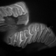
(Click on image for MOV file, or here for AVI file.) |
Venom duct of cone snail (Conus magus)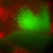
(Click on image for MOV file, or here for AVI file.) |
| 60x/1.1NA (water), rhodamine auto-fluorescence. | Radular sac, 20x/0.5NA (water), red image is transmitted light, green is rhodamine auto-fluorescence in the rhodamine band. |