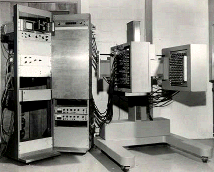
The logical extension of positron instrumentation was a design using two 2-dimensional arrays. PC-I was the first instrument using this concept and was designed in 1968, completed in 1969 and reported in 1972 [11] [20]. The first applications of PC-I in tomographic mode as distinguished from the computed tomographic mode were reported in 1970 (Brownell et al 1970 [10]). PC-I incorporated rotation and translation of the two detectorbanks and included interpolative motion of the detectors to improve sampling and image quality. PC-I could produce images on planes parallel to the detector planes or tomographic images on planes within the object. PC-I was not patented because it was disclosed in several papers but was covered by a US Atomic Energy Commission Record of Invention (S-40, 757) with date of inception as June 1968, completion in 1969 and first tests in May 1971 (Figure 4).
The original intent was to use PC-I to obtain focused images on planes parallel to the detector planes and tomographic images on transverse planes. The use of PC-I to obtain computed tomographic images or PET images evolved over this period of time.
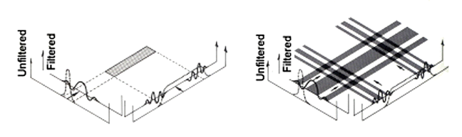
In early 1970, David Chesler in our group at MGH conceived of filtered back projection. In the summer of 1970 he tested filtered back projection, including the effects of Poisson noise, by computer simulation. On Veterans Day of 1970 he collected both emission and transmission data with a `bench top scanner'. From this data he was able to produce three types of computed tomographic images: an emission image, a transmission image and an absorption-corrected emission image. Figure 5 illustrates the concept of filtered back projection and was presented by Chesler at the Meeting on Tomographic Imaging in Nuclear Medicine September 15-16, 1972. This development of filtered back projection was the first reconstruction of this type to be applied to PET and CT data. (Chesler 1971 [24], Chesler 1973 [25] (Figure 6) and Chesler et al 1973 [26] (Figure 7).) The original intent was to use PC-I to obtain focused images on planes parallel to the detector planes and tomographic images on transverse planes. The filtered back projection algorithm was immediately applied to data from PC-I and the subsequent computed tomographic images were dubbed PET images as an acronym for positron emission tomography. (Brownell and Burnham 1972 [11], Brownell and Burnham 1973 [12], and Brownell et al 1978 [16]). The excellent book by Steve Webb entitled ``From the Watching of Shadows" [52] gives a well documented account of the development of X-ray CT as well as the early work in positron emission tomography (PET) and single photon computed emission tomography (SPECT) [39] [40]. Two names stand out in CT, Godfrey Houndsfield (Hounsfield 1973 [35]) and Allan Cormack (Cormack 1973 [29]), both of whom were recognized for their contributions by sharing the Nobel Prize. (Hounsfield used an iterative technique on X-ray CT data.) Houndsfield obtained his first patent in August of 1972 well after the first clinical trials at the Atkinson Morley Hospital in October 1971 (Ambrose 1973 [2]).
 |
 |
 |